Fascia Insights: Diversity in Structure and Function
Grafics for Download
All the illustrations offered here have been compiled with a lot of research, tireless enthusiasm and the friendly support of Robert Schleip from the Fascia Illustration Research Group.
These graphics serve to illustrate the complex and interactive tasks of the fascia system for research, medicine and sports. For the first time, they show important sub-areas of this exciting fabric in detail, vividly and cutting-edge.
We would like to thank the Fascia Research Society for the presentation at the Fascia Research Congress 2022 as well as the fascia luminaries Gary Carter, Peter Friedl, Stuart McGill, J.C. Guimberteau, Werner Klingler, Helene Langevin, Tom Myers, Yuval Rinkevich, Carla Stecco and Jan Wilke for their time, hints and recognition.
Miriam Wessels, Heike Oellerich and Juliane Galke
⇒ Fascia illustration: Fascial Continuity of the Functional Levels
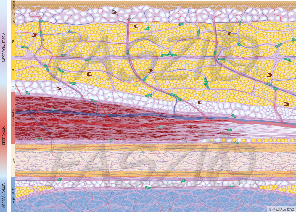
This fascia graphic shows the three fascial functional levels (superficial, deep and visceral plane) that are formed and connected within the fascia network. Due to the fascial structural diversity and continuity, each level acts independently and at the same time remains part of the three-dimensional fascia network with body-wide interaction. The loose fascia tissue (LCT) allows all structures to slide against each other, thus enabling interaction, transfer and mobility within and between the planes. Connective tissue strands (retinacula cutis) anchor deeper, firmer structures (periosteum, myofascia) to the skin and thus limit the radius of displacement. Its fibers first run slightly diagonally through the deep adipose tissue (DAT), then meet the superficial fascia and from there move in a straight line through the superficial adipose tissue (SAT) to the dermis.
All together they create a functional symbiosis of flexibility and stability.
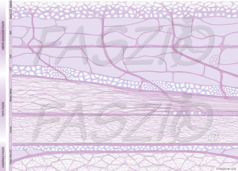
⇒ Fascia illustration: Fascial Continuity of the Fascia
This fascia graphic shows the continuity of the three-dimensional fascia network. In contrast to the above illustration, blood vessels, muscle, fat, connective tissue cells, etc. have been removed, so that only the fascial fiber network can be seen. This impressively illustrates the continuous connecting structure of the fascial tissue and its mutability.
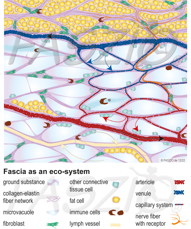
This fascia graphic shows the structure of the loose fascia tissue (LCT). The multiple interactions of cells, metabolism, immune system and nerve functions with and within this fluid-rich fascia structure are clearly visible.
The fibrous network, which is moistened with basic substance, forms arterial and venous vessels, lymphatic vessels and serves as an embedding for nerve fibers. It is the place of residence, transport route as well as supply and disposal environment of and for cells (including immune, fat, nerve, connective tissue cells). In addition, special cell construction workers (fibroblasts) operate there, who are responsible for building up and breaking down the tissue network.
With the help of a vacuole architecture, water is stored and a lubrication, deformability and stabilization of the tissue is made possible.
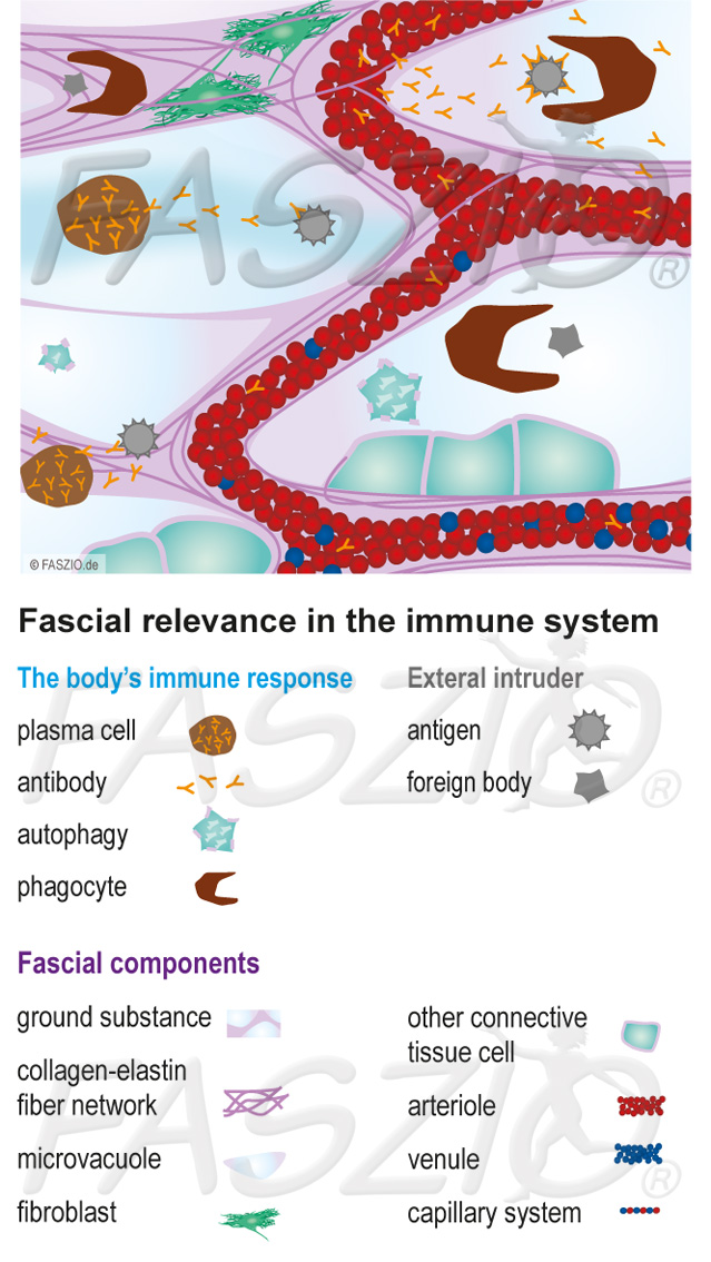
⇒ Fascia illustration: Fascial relevance in the immune system
This fascia graphic shows the dependence of the immune system on the fascia. The undisturbed fascial transport and supply property is the regulatory prerequisite for a well-functioning immune response as well as for cell recycling (autophagy) by recognizing, breaking down and recycling the cell’s own components.
Within the permeable basic substance, immune cells fight invading foreign bodies and antigens. Overall, the surface tension of the bound water forms a protective barrier to make it more difficult for it to penetrate.
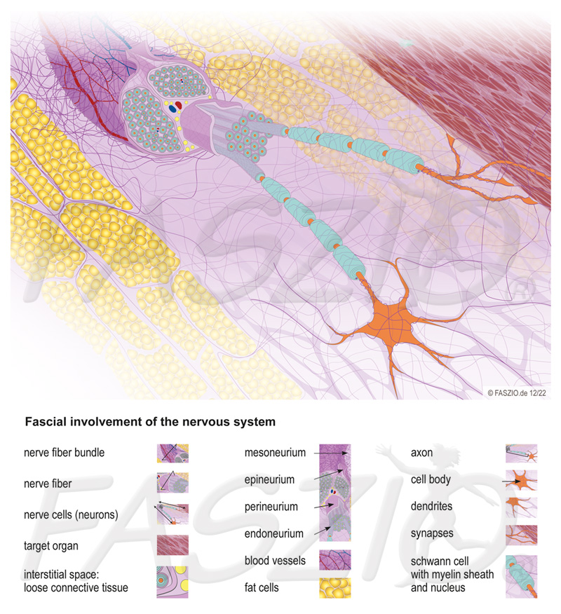
⇒ Fascia illustration: Fascial involvement of the nervous system
This graph shows how closely the nerve and its ability to function are connected to the fascia. The fascial tissue forms the nerve bed, runs through and supplies the nerve and connects it with its surroundings.
The receptors are stretched in the fibrous network and receive stimuli, the transmission of which via the axons is also influenced by the quality and lubrication of the fascia. This controls, among other things, the sensation, the ability to react and the handling of stress.
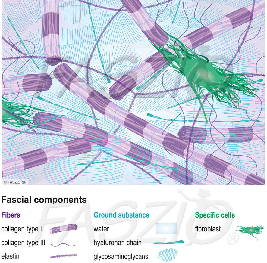
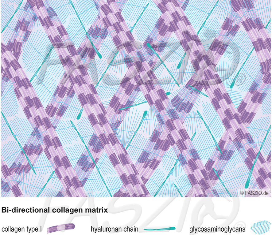
This fascia graphic concentrates on the generally relevant components and mainly uses simplifying generic terms.
The fascia is a network of tissue made up of collagen fibers (tensile and tear-resistant, can spring back like a catapult) and elastin fibers (loose compound that can elongate up to 150% reversibly). The fibres are connected via sugar-protein compounds, which in turn bind water (basic substance).
The fibroblasts belonging to the fascia produce fibrous elements and connecting proteins. They are responsible for permanent assembly, dismantling and conversion.
As a result, the fascial tissue is able to actively adapt to the respective requirements.
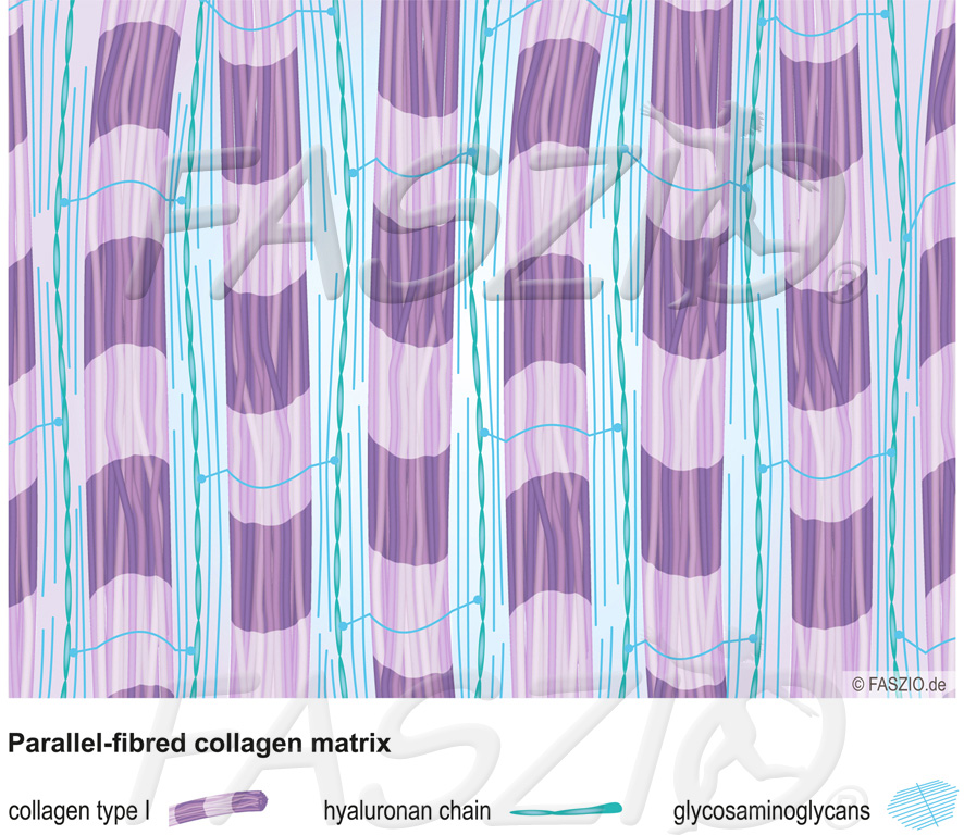
⇒ Fascia illustration: Parallel-fibred collagen matrix
This fascia graphic shows the parallel course of collagen type I fibers, which is found in tissue structures that require strong tensile strength (e.g. tendons, ligaments). Various connecting proteins provide the arrangement, fluidity and functionality.
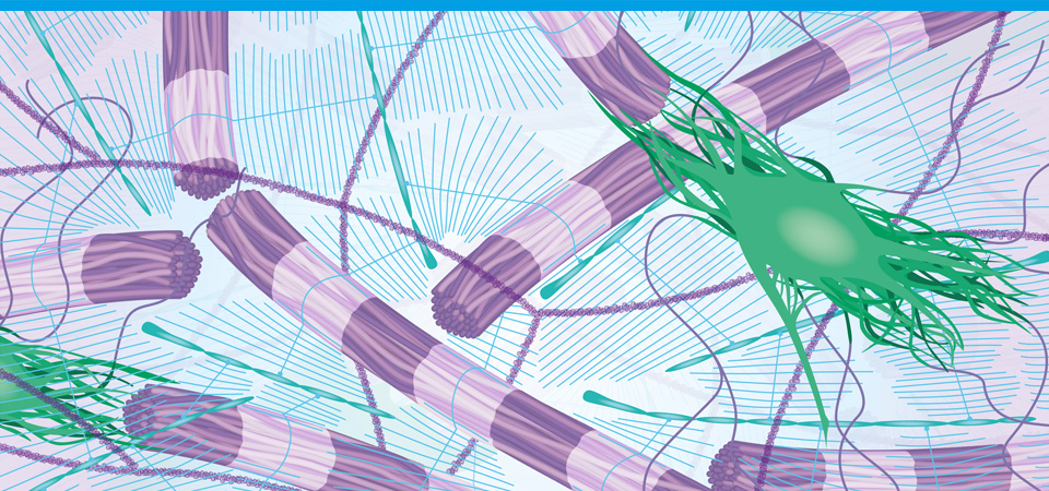
This fascia graphic shows the bi-directional course of collagen type I fibers found in tissue structures with multidirectional radius of motion and volume changes (e.g. muscle bodies, organ sheath). Connecting proteins ensure the arrangement, fluidity and functionality.
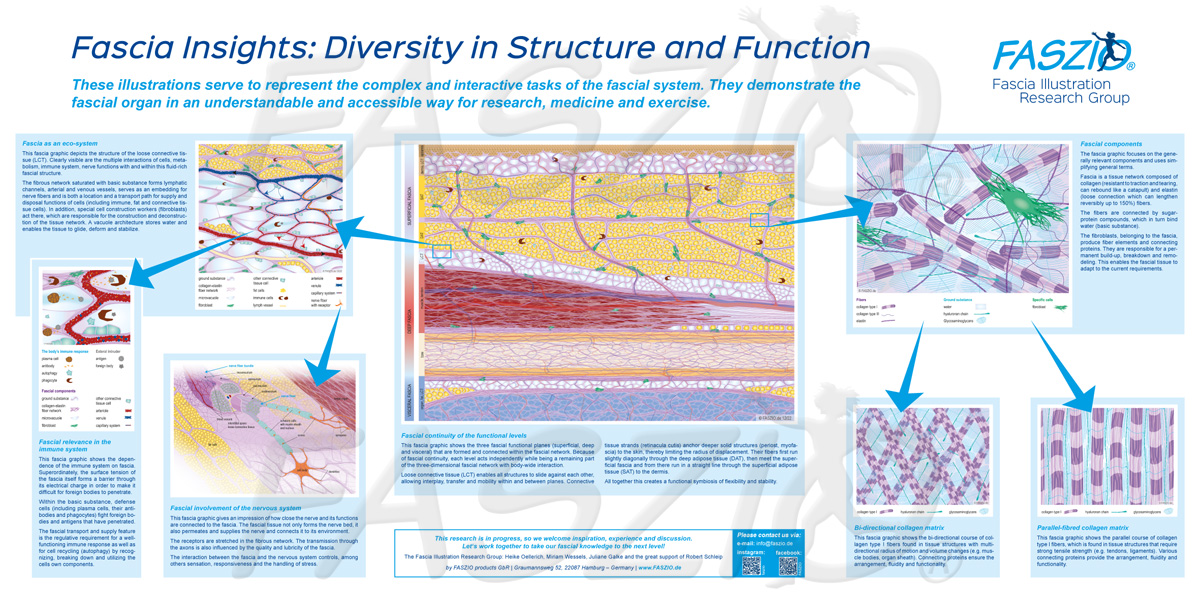
⇒ Fascia Poster: Fascia Insights – Diversity in Structure and Function
This compilation shows all the graphics at a glance to illustrate the complex and interactive tasks of the fascia system. They show the fascia organ in an understandable and accessible way for research, medicine and sports.